 Living Beings
Compare species
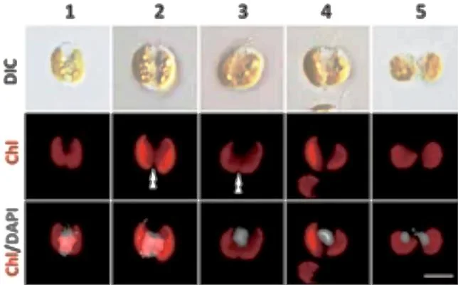
Guillardia theta
Guillardia theta
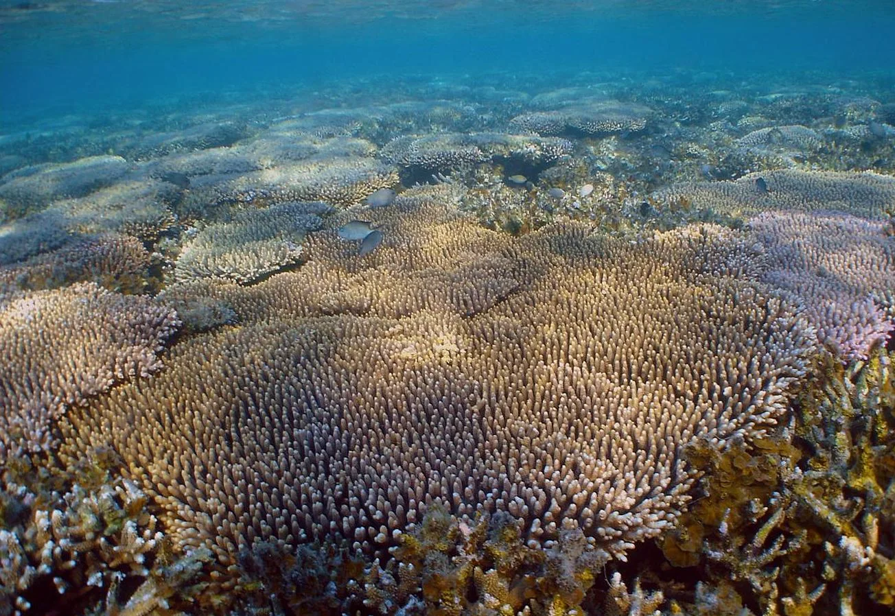
Acropora millepora
Acropora millepora
How to compare two organisms
Choose two species and trace their closest shared group. Then compare a trait with the same definition on each side. Check the units, life stage and context before interpreting a measurement or range.
Missing data means that the collection lacks a sourced value. Where a characteristic does not apply to a lineage, the comparison labels it separately. Open the two profiles for context and follow their sources before drawing a conclusion.
Evidence from the profiles
Compare their characteristics
| Trait | Guillardia theta | Acropora millepora |
|---|---|---|
| Physical traits | The microscopy montage combines transmitted-light images with fluorescence channels. The colored signals mark cellular structures and should not be mistaken for the organism’s natural visible coloration. | Colonies form branching structures covered by small skeletal cups that house polyps. Colony color alone is insufficient for secure species identification. |
| Habitat and range | The cited sequencing studies describe laboratory strains. A complete field distribution and habitat preference should not be inferred from those cultures alone. | Shallow, well-lit marine reef environments support the coral and its photosynthetic partners. |
| Diet and nutrition | A chloroplast supports photosynthesis. The plastid contains its own compact genome, distinct from the main nuclear genome. | Polyps capture small suspended food, while algal symbionts contribute photosynthetic products. |
| Behavior and activity | Cellular research distinguishes nuclear, plastid and mitochondrial functions rather than assigning all activity to a single genome. | Tentacles and mucus help the colony interact with surrounding seawater. Recognition mechanisms influence responses to microbes. |
| Reproduction and life cycle | Cells grow and divide as microscopic eukaryotes. The image follows cellular stages; these are not separate species or animal life stages. | Colonies grow by producing additional polyps. Sexual reproduction can release gametes that develop into settling larvae. |
| Geographic distribution | Data unavailable | The species occurs in the Indo-Pacific reef region. Species-level locality records should accompany detailed range mapping. |
| Ecological role | As an alga it contributes a photosynthetic lineage to microbial diversity. The cited genome studies do not provide a full account of its abundance in nature. | Skeletal growth contributes reef structure and habitat. Symbiotic interactions are central to energy supply and colony health. |
| Distinctive adaptations | A nucleomorph, a highly reduced remnant nucleus of the engulfed red-algal partner, persists beside the plastid. This is evidence of secondary endosymbiosis. | A colonial body and partnerships with photosynthetic cells support growth in well-lit reef environments. |
| Conservation | Data unavailable | Thermal stress, bleaching and degradation of reef habitat are concerns. A source on molecular recognition does not establish a current Red List category. |
Classification
Shared ancestry
Closest shared taxon: Eukaryota.