 Living Beings
 Framed for display
Compare species
Escherichia coli
Escherichia coli
Haloferax volcanii
Haloferax volcanii
How to compare two organisms
Choose two species and trace their closest shared group. Then compare a trait with the same definition on each side. Check the units, life stage and context before interpreting a measurement or range.
Missing data means that the collection lacks a sourced value. Where a characteristic does not apply to a lineage, the comparison labels it separately. Open the two profiles for context and follow their sources before drawing a conclusion.
Biological comparison
Means, ranges and reported maxima describe different things. Check the source and population before comparing values.
Classification
Shared ancestry
Shared navigation root · Life.
Separate lineages
| Characteristic | Escherichia coli | Haloferax volcanii |
|---|---|---|
Scientific measurements | Scientific measurements | |
| No reference measurements are available for this pair. | ||
Context-specific measurements | Context-specific measurements | |
| Cell length |
1.94–2.72 µm
Reported range · Study strain E. coli B · Laboratory study · Context-specific record Source and measurement contextReported range across sample mean cell lengths in a laboratory study of a strain of E. coli B. The endpoints do not define the range of individual cells or of the species. Length of a single cell under the reported conditions.
|
1–3 µm
Reported range · DS2 reference account · Context-specific record Source and measurement contextLength listed in the study’s phenotypic summary. Cell shape varies. Length of a single cell under the reported conditions.
|
Natural history | Natural history | |
| Nutrition and energy acquisition |
No separate account recorded |
Organic nutrients can support growth in simple defined media. Salt balance is a physiological requirement rather than a food source. Source 1 ↗Source 2 ↗ |
| Habitat |
E. coli can occur in intestinal tracts, water, food and other environments. Water monitoring uses counts of E. coli as …E. coli can occur in intestinal tracts, water, food and other environments. Water monitoring uses counts of E. coli as an indicator of fecal contamination. An indicator result describes conditions in a sampled body of water and cannot by itself identify a disease-causing strain or the origin of the contamination. |
Saline environments and appropriately salt-rich culture media support growth. Source 1 ↗Source 2 ↗ |
| Geographic distribution |
No separate account recorded |
The type strain DS2 is a defined reference for genomic work. Worldwide use in laboratories is separate from the distribution of environmental populations. Source 1 ↗Source 2 ↗ |
| Morphology |
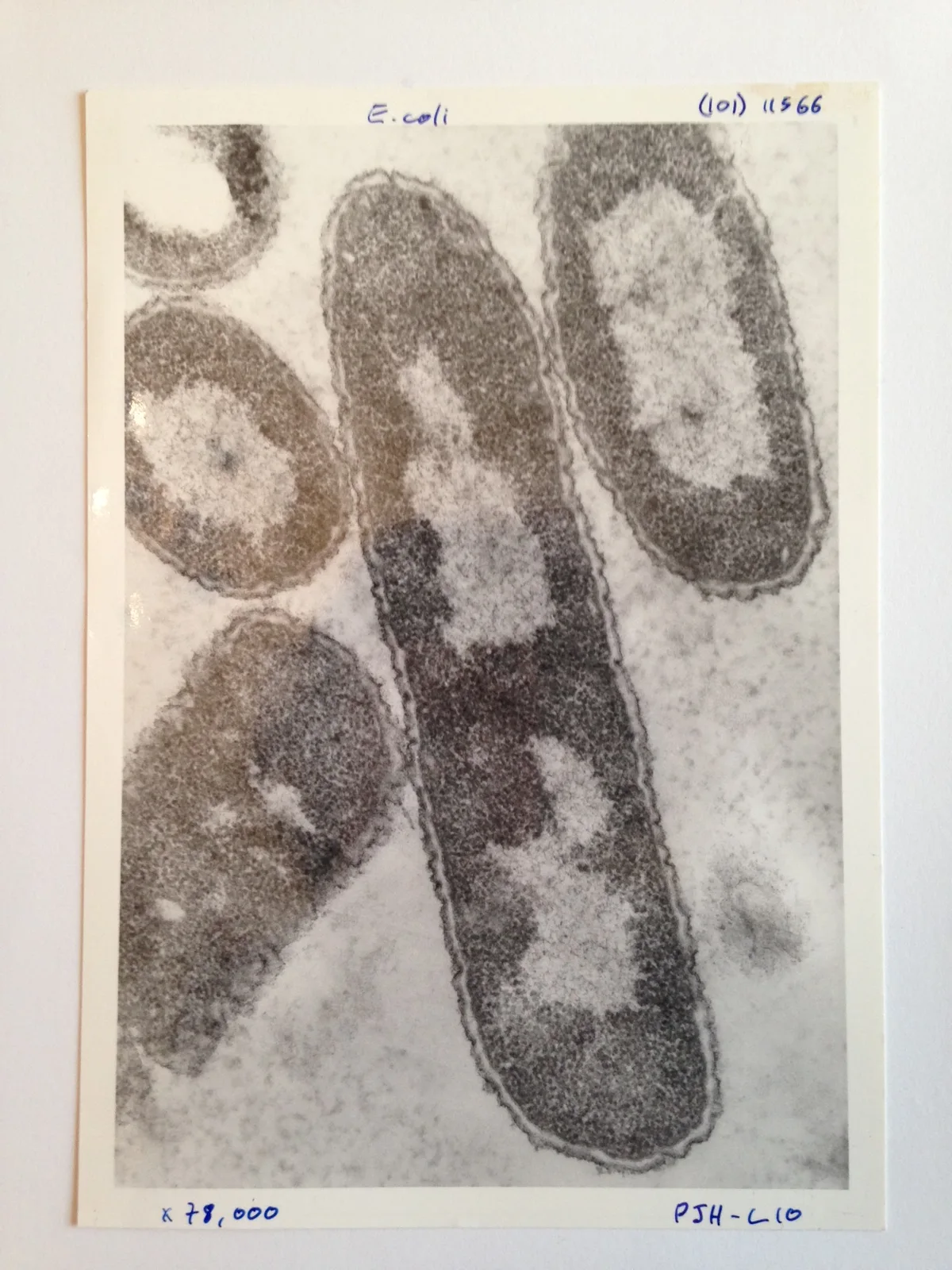
The species name refers to a group of bacteria whose strains cannot be distinguished by ordinary rod shape alone. A micrograph is useful for seeing cell form, but strain identity and gene content require other evidence. Source 1 ↗ |
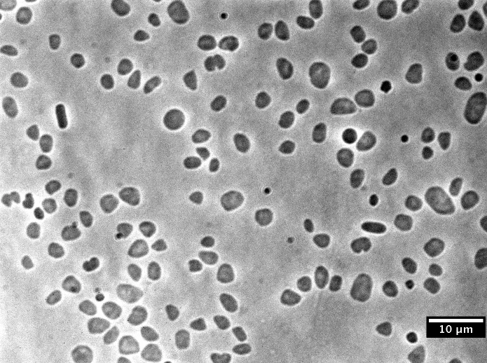
Cells require microscopy for observation. The sequenced type strain DS2 carries a main chromosome, three smaller chromosomes and a plasmid. …Cells require microscopy for observation. The sequenced type strain DS2 carries a main chromosome, three smaller chromosomes and a plasmid. Those DNA elements are not membrane-bound nuclei. Their organization helps explain why studying an archaeon involves more than treating it as a small version of a familiar bacterium. |
| Reproduction and life cycle |
No separate account recorded |
Cells multiply through division. Experiments can follow inherited genetic changes through successive generations of a culture. Source 1 ↗Source 2 ↗ |
| Ecological role |
Some E. coli types occur as ordinary members of intestinal microbial communities. Others carry traits associated with illness. The presence …Some E. coli types occur as ordinary members of intestinal microbial communities. Others carry traits associated with illness. The presence of the species name in a sample does not by itself identify the strain or establish that it caused disease. |
Its life in saline microbial communities provides a contrast with the marine ammonia oxidizers and gut methanogens elsewhere in Archaea. Source 1 ↗Source 2 ↗ |
| Behavior and activity |
No separate account recorded |
Cellular responses to salt and available resources are studied using a developed genetic toolkit. Source 1 ↗Source 2 ↗ |
| Environmental adaptations |
A genome comparison between an O157:H7 strain and a laboratory K-12 strain found shared sequence alongside regions specific to each …A genome comparison between an O157:H7 strain and a laboratory K-12 strain found shared sequence alongside regions specific to each lineage. The shared part helps explain why they are related. The differences help explain why observations about one strain should be reported with its strain name instead of being assigned to every E. coli cell. |
Salt-adapted physiology and a multipartite genome are important features of the reference strain. Source 1 ↗Source 2 ↗ |
| Conservation |
No separate account recorded |
No separate account recorded |
Sources from both profiles
Escherichia coli
More comparisons
Framed for display
